

Figure 1
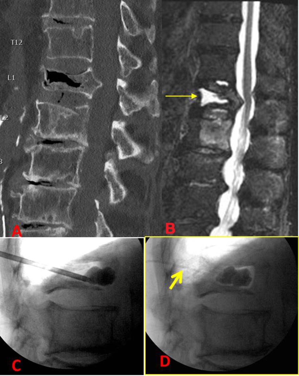
Figure 1
(A) Computed tomography (CT) sagittal image showing the cleft in the L1 vertebrae. (B) Magnetic resonance imaging (MRI) showing fluid-filled lytic cleft (thin yellow arrow). (C) Cement mass stuck to the trocar. (D) Thick Yellow arrow showing the cement tail. The cement mass moved dorsally with removal of the trocar.
In this issue


Related Articles
Cited By...
- No citing articles found.