


Abstract
Background Nuclear imaging modalities are increasingly advancing spinal diagnostics. This study evaluates the prevalence of high uptake in bone scan and single photon emission computed tomography combined with high-resolution computed tomography (SPECT/CT) in the spine and sacroiliac joint (SIJ) and compares the diagnostic performance of BS to SPECT/CT in detecting metabolic activity linked to neck and back pain.
Objective The primary objective was to assess the sensitivity, specificity, and diagnostic accuracy of BSs compared with SPECT/CT for spine and SIJ evaluation.
Methods This retrospective study evaluated data from patients with spinal complaints who underwent spine-focused SPECT/CT alongside whole-body BS at a tertiary institution.
Results A total of 110 patients were included, with 48 cervical spine, 34 thoracic spine, and 91 lumbar spine and SIJ SPECT/CT scans. For the cervical spine, BS sensitivity, specificity, and accuracy were 41.5%, 100%, and 50%, respectively. For the thoracic spine, these values were 50.0%, 100%, and 73.5%, respectively. For the lumbar spine, they were 72.9%, 100%, and 79.1%. For the SIJ, sensitivity, specificity, and accuracy were 38.2%, 96.5%, and 74.7%, respectively.
Conclusions Bone scans demonstrated reasonable sensitivity and high specificity, particularly for lumbar spine and SIJ evaluation, making them a useful screening tool in resource-constrained settings. However, SPECT/CT showed superior performance in detecting osteometabolic activity and provided more detailed functional and structural insights for diagnosing and managing degenerative spinal conditions.
Clinical Relevance This study highlights the potential complementary role of bone scans in spinal diagnostics.
Level of Evidence 3.
- nuclear imaging
- neck pain
- back pain
- SPECT/CT
- bone scan
- diagnostic performance
- prevalence
- degenerative spine
- sacroiliac joint
Introduction
Neck and back pain are major global public health concerns, with high incidence rates and a substantial impact on disability levels worldwide.1,2 Among the various contributing factors to neck pain, degenerative changes such as osteoarthritis stand out as prominent causes.3 However, correlating specific symptoms with distinct subtypes of spinal degenerative changes remains a challenging task.4
A variety of imaging modalities are available for diagnostic evaluation, including x-ray, computed tomography (CT), and magnetic resonance imaging.5,6 Recently, the use of nuclear diagnostic methods such as whole-body 2-dimensional bone scan (BS) and 3-dimensional single photon emission CT combined with high-resolution CT (SPECT/CT) has been increasing. SPECT/CT, as a hybrid imaging technique, provides cross-sectional images that reveal osteoblastic activity, offering valuable functional insights in addition to structural details, particularly in regions of interest.7–9
This study aimed to examine the prevalence of high uptake in BS and SPECT/CT in the spine and sacroiliac joint (SIJ) and to evaluate the diagnostic performance and accuracy of these imaging techniques.
Methods
Study Design, Population, and Analysis Sets
This retrospective analysis utilized data from patients presenting with back complaints at the spine unit of a tertiary institution. The study included individuals who underwent spine-focused SPECT/CT between January 2019 and January 2023. For all patients, a whole-body BS was performed, providing imaging of the entire spine, while SPECT/CT focused on a specific region of interest determined by clinical suspicion.
The primary objective was to analyze findings in designated spinal regions and the SIJ. The dataset was cross-sectional and included only patients who had provided general consent for the use of their data. Exclusion criteria included the presence of infections, age younger than 18 years, and presence of known tumors.
This approach allowed for a targeted evaluation of the diagnostic performance of SPECT/CT in conjunction with BS for specific spinal and SIJ regions while adhering to ethical and clinical standards.
Primary Outcome and Endpoints
The primary outcome was to evaluate the diagnostic characteristics, including sensitivity, specificity, and accuracy, for detecting spinal and SIJ findings. These measures were assessed by comparing results from BS and SPECT/CT.1–3
Both endpoints were binary in nature (i.e., either absent or present). The specific binary endpoints evaluated were the following:
Spots of high uptake on BS.
Spots of high uptake on SPECT/CT.
Data were extracted from patient records and radiologists’ reports of BS and SPECT/CT findings. A patient was classified as positive if there was any indication of metabolic active degenerative disease (eg, facet joint or SIJ arthritis and osteochondrosis) on BS or SPECT/CT. Conversely, a negative classification was assigned if no high uptake was detected on either imaging modality.
Figures 1 and 2 provide illustrative examples of positive findings observed in the patient group studied. These cases demonstrate the imaging characteristics of high uptake regions indicative of degenerative processes.

Forty-five-year-old male patient with pain in the thoracic spine region. The bone scan (bottom left) and the single photon emission computed tomography (CT) combined with high-resolution CT (top images) show high uptake in the facet joints of T7 to T10 bilaterally. The native CT (bottom right) reveals facet joint osteoarthritis.

Seventy-eight-year-old female patient with lower back pain. The bone scan (top image) and the single photon emission computed tomography (CT) combined with high-resolution CT (bottom left) show high uptake in the right sacroiliac joint (SIJ). The native CT (bottom right) reveals SIJ osteoarthritis.
Statistical Analysis
All analyses were performed in R version 4.3.1. This study is explorative and descriptive in nature. No P values are reported. Summary statistics of patient characteristics were presented for all patients using frequencies and percentages for categorical variables and either means and SDs or medians and interquartile ranges for numerical variables. The proportion of patients with uptake on BS and SPECT/CT was described per region of interest.
Analysis of the Primary Objective
Our primary focus was to assess the correlation between findings from each diagnostic tool. We presented a contingency table comparing diagnoses from both BS and SPECT/CT to illustrate the agreement and disagreement between the methods. The diagnostic performance parameters—sensitivity, specificity, and accuracy (the proportion of correctly classified patients)—were measured alongside their respective 95% Wilson confidence intervals.
Additional Variable Definitions
To evaluate the diagnostic measures, we dichotomized radiological diagnosis as yes or no for the following 3 occasions according to the clinical routine:
Facet joint osteoarthritis (yes/no).
Osteochondrosis (yes/no).
Both diagnosis (yes/no).
Results
A total of 110 patients were included in the analysis, comprising 45 men (41%) and 65 women (59%). Imaging studies included 48 SPECT/CT scans of the cervical spine, 34 of the thoracic spine, and 91 of the lumbar spine and SIJ. BS was available for all cases (n = 110, 100%).
Prevalence of High Uptake on BS and SPECT
Cervical Spine
Of the 110 BS scans, 22 (20.0%) demonstrated high uptake in the cervical spine. In comparison, 41 (85.4%) of the 48 cervical SPECT/CT scans showed high uptake.
Thoracic Spine
Among the 110 BS scans, 18 (16.4%) showed high uptake in the thoracic spine. In contrast, 18 (52.9%) of the 34 thoracic SPECT/CT scans exhibited high uptake.
Lumbar Spine
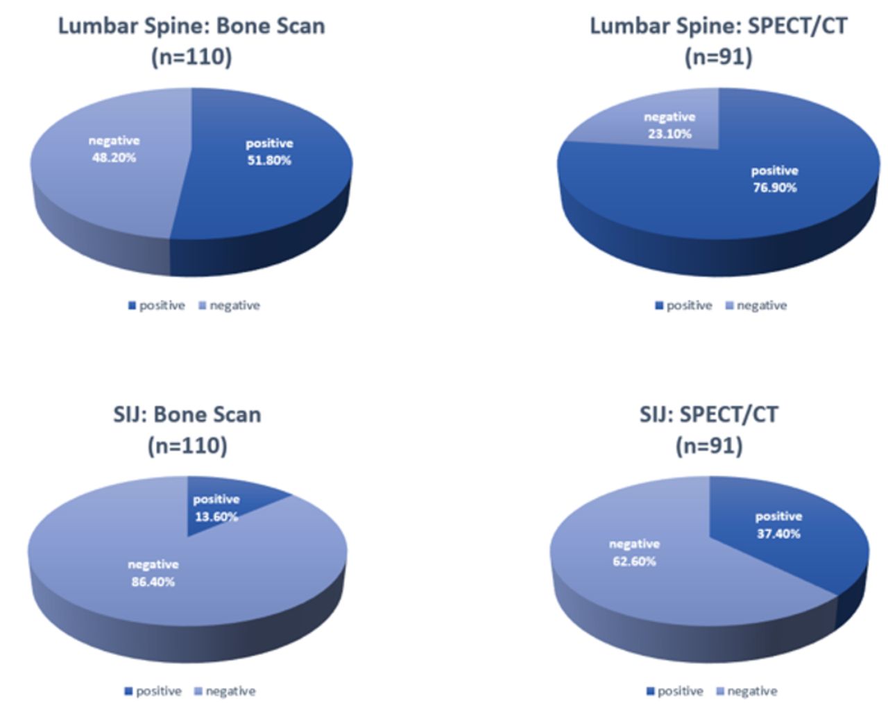
Of the 110 BS scans, 57 (51.8%) demonstrated high uptake in the lumbar spine. Meanwhile, 70 (76.9%) of the 91 lumbar SPECT/CT scans revealed high uptake (Figure 3).

The diagrams illustrate the prevalence of the high uptake in the specific regions of interest in bone scan vs SPECT/CT. Abbreviations: CT, computed tomography; SPECT/CT, single photon emission computed tomography combined with high-resolution CT.
Sacroiliac Joint
Among the 110 BS scans, 15 (13.6%) exhibited high uptake in the SIJ. In contrast, 34 (37.4%) of the 91 SIJ SPECT/CT scans showed high uptake (Figure 3).
Diagnostic Performance of BS Compared With SPECT/CT
The Table presents the sensitivity, specificity, and diagnostic accuracy of BS compared with SPECT/CT for each region. Elevated uptakes identified on BS were consistently observed on SPECT/CT, but only about half of the uptakes detected on SPECT/CT were visible on BS alone.
Sensitivity, specificity, and diagnostic accuracy of BS compared with SPECT.
Cervical Spine
Among 48 cervical SPECT/CT scans, 41 were positive and 7 were negative. Of these, 17 patients had both positive BS and SPECT/CT results, while 24 SPECT-positive patients had negative BS results. All SPECT-negative patients also had negative BS results. The sensitivity of BS was 41.5%, specificity was 100%, and diagnostic accuracy was 50%.
Thoracic Spine
A total of 34 thoracic SPECT/CT scans were analyzed, with 18 positive and 16 negative results. Nine patients had positive results on both BS and SPECT/CT, while 9 SPECT-positive patients had negative BS results. All SPECT-negative patients also had negative BS results. The sensitivity, specificity, and accuracy of BS were 50.0%, 100%, and 73.5%, respectively.
Lumbar Spine
Of the 91 lumbar SPECT/CT scans, 70 were positive, and 21 were negative. Among these, 51 patients had both positive BS and SPECT/CT results, while 19 SPECT-positive patients had negative BS results. All SPECT-negative patients also had negative BS results. The sensitivity, specificity, and accuracy of BS were 72.9%, 100%, and 79.1%.
Sacroiliac Joint
Out of 91 SIJ SPECT/CT scans, 34 were positive, and 57 were negative. Thirteen patients had both positive BS and SPECT/CT results, while 21 SPECT-positive patients had negative BS results. All SPECT-negative patients also had negative BS results. The sensitivity, specificity, and accuracy of BS for the SIJ were 38.2%, 96.5%, and 74.7%.
Discussion
Neck and back pain, often linked to degenerative changes like osteoarthritis, remains a clinical challenge. The global burden of neck pain underscores its widespread prevalence and associated disability.2 These conditions impose significant economic and health care challenges globally.1
Advances in nuclear imaging modalities have improved the diagnostic process for spinal degeneration.5,7 In particular, SPECT/CT has demonstrated high sensitivity in identifying pain generators, such as facet joint arthritis, in patients with chronic back and neck pain.7–9 Over the past 2 decades, SPECT/CT has evolved into a mature technology, supported by a robust body of evidence.10 Our study provides further insight into the diagnostic accuracy of BS compared with SPECT/CT in identifying metabolic activity associated with degenerative changes. Our results highlight the potential of utilizing whole-body BS, initially performed for various clinical indications, to assess spinal degeneration. Moreover, it is the first to provide insights into the diagnostic performance of BS compared with the more advanced nuclear imaging modality, SPECT/CT.
For the cervical spine, BS sensitivity was relatively low (41.5%), reflecting its limited ability to detect metabolic activity in this region. These findings align with previous research, which highlighted the challenges of correlating clinical symptoms with imaging findings in cervical degeneration.4 However, the high specificity (100%) supports the use of BS as a confirmatory tool in cases where SPECT/CT is unavailable.
In the lumbar spine, BS showed greater sensitivity (72.9%) and diagnostic accuracy (79.1%), making it a viable screening tool in cases where BS is already available from previous nuclear imaging studies conducted for other indications.
The findings of our study are consistent with those reported by Holder et al, who observed a sensitivity of 100% for SPECT/CT and 71% for BS.11 While their work primarily focused on planar and high-resolution SPECT/CT bone imaging in the context of wrist diagnostics, our study uniquely applies these comparisons to spinal degeneration, specifically facet joint arthritis. This offers valuable insights into the diagnostic utility of BS and SPECT/CT in the spine.
These findings highlight the complementary strengths of each modality. SPECT/CT remains a preferred technique for diagnosing osteometabolic activity due to its superior sensitivity and precise localization of abnormalities.⁷ However, the high specificity of BS across all regions (100%) enhances its utility as a practical alternative in resource-limited settings or when SPECT/CT is unavailable. BS also serves as a valuable complementary tool when paired with other diagnostic modalities such as magnetic resonance imaging or radiography. Future research should focus on larger, more diverse populations to validate these findings and explore optimal strategies for integrating BS and SPECT/CT into clinical practice.
Limitations
This study has some limitations. Its single-center design may influence generalizability, particularly for patients outside a tertiary care setting. While recruitment from a specialized institution may introduce selection bias, it ensures consistent imaging quality. The exclusion of patients with infections or tumors helped focus the sample but may limit broader applicability. The retrospective nature of the study, while effective for comparing imaging modalities, relies on existing records, which may vary in detail. Future studies incorporating multicenter designs and stronger clinical correlations could further validate these findings and enhance their applicability. Despite these considerations, the study offers valuable insights into the diagnostic utility of BS and SPECT/CT.
Conclusion
BSs demonstrated reasonable sensitivity and high specificity, particularly for lumbar spine and SIJ evaluation, making them a practical screening tool in resource-constrained settings. While SPECT/CT has demonstrated superiority due to its enhanced performance in detecting osteometabolic activity and providing critical functional and structural insights, BSs play a complementary role in spinal diagnostics. These findings support the integration of BS as a reliable option for diagnosing and managing degenerative spinal conditions, particularly when access to SPECT/CT of the spinal region of interest is not available.
Acknowledgements
We would like to thank the research department of the cantonal hospital Aarau for their support.
Footnotes
Funding Financial support has been granted by the research department of our institution (Aarau Cantonal Hospital).
Declaration of Conflicting Interests The authors report no conflicts of interest in this work.
Ethics Approval Ethics Approval 2023-00526 (Ethics Committee Northwest Switzerland).
Data Availability Statement The data supporting the findings of this study can be obtained from the corresponding author upon reasonable request. Access to patient data is governed by ethical and privacy considerations in accordance with institutional guideline.
- This manuscript is generously published free of charge by ISASS, the International Society for the Advancement of Spine Surgery. Copyright © 2025 ISASS. To see more or order reprints or permissions, see http://ijssurgery.com.
In this issue


Jump to section
Related Articles
Cited By...
- No citing articles found.